- Contact Us
- |
- Sitemap
- |
- Home
Follow us on :


The opacification of the normal transparent lens is called cataract. The Latin word 'cataracta' means 'waterfall'. Imagine trying to peer through a sheet of falling water or through a frosted or fogged-up window.
The earliest surgery treatment began in India. It was known as 'couching'. The sclera
was incised. Then the lens was dislocated backward into the vitreous and out of
optical axis.
This procedure was performed for more than two thousand years until the mid-eighteenth
century. Great progress in cataract surgery has been made in recent years with the
introduction of micro-surgical instruments, microscope and modern surgical techniques
like phaco-emulsification. In the early stages of cataract development, all that
is needed to correct your vision with glasses is a change in prescription. As the
cataract develops and begins to affect your lifestyle, it needs to be removed. Cataract
surgery, the most commonly performed operation, is safe and effective in 95% patients
with enhancement in vision.
The primary function of the bi-convex lens is to refract and focus light on the
retina while remaining transparent. This transparency depends on the maintenance
of structural (anatomic) & functional (physiologic) integrity.
The lens is 66% water,the least hydrated organ of the body. The remaining bulk is
composed mainly of protein. It is devoid of any blood supply and derives its nourishment
from the surrounding aqueous and vitreous.
Cataracts are classified as per their morphology (form and structure) and as per
their maturity.
The cataract occurs as a result of the natural aging process of lens fibres which become opaque over a period of time.
Steroid-induced cataract :
This occurs as a result of excess intake of oral steroid or putting steroid drops
in the eye.Drugs induced: Chlorpromazine, Miotics, Busulphan, Amiodarone, gold
Secondary cataract:
Here, cataract develops as a result of some other primary ocular disease such as
chronic inflammation and glaucoma.
One may not be aware that a cataract is developing if the size and location of the cloudy areas in the lens are not in the pupillary area. As the cataract progresses, there is deterioration of distant and near vision.
It varies from person to person but as a general rule, most cataracts develop slowly over a period of time. A cataract can take months or even years to reach a point where it adversely affects vision.
Visual acuity :
Checking vision of both eyes unaided and aided with glasses and pin-hole vision
to know the improvement as well as to get the general idea about the macular function
of the eyes. This will help in prognostic evaluation of visual recovery after cataract
surgery.
Intra ocular pressure :
If intra-ocular pressure increases as a secondary to cataract, surgery is needed
to prevent further complications.
Slit-lamp examination :
To know the type of cataract along with its opacity, morphology and etiology or
any associated ocular pathology.
Direct and indirect ophthalmoscopy :
Dense opacity (cataract will prevent retinal evaluation)
A-scan biometry:
To calculate the AL and IOL power for implantation In cases of mature cataract the
posterior segment of the eye is evaluated.
No presently available medical treatment can prevent cataracts or reverse them once they develop.
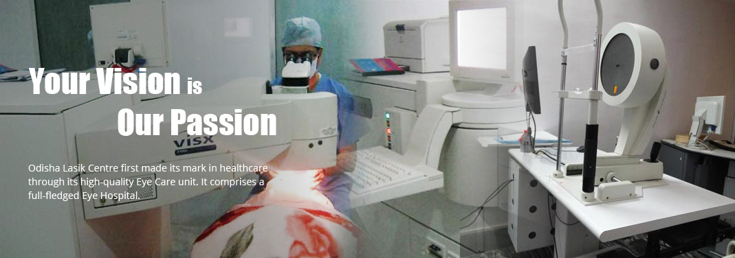
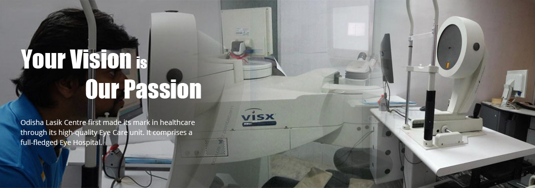
Phaco-emulsification is the latest technology in cataract surgery. It is a micro-incision stitch-less operation, where the cataract is emulsified by ultrasound energy, liquefied and sucked through the phaco-emulsifier probe. Then a foldable intra-ocular lens is implanted in the eye permanently.
It is the least traumatic form of cataract surgery with early rehabilitation and recuperation. Vision restoration is possible in a short period of time.
During cataract surgery, the natural lens of the eye that has turned opaque is removed, resulting in loss of focusing power of the eye. This situation would be parallel to clicking a photograph without a camera lens-the picture would be extremely blurred. When the natural lens of the eye is removed an artificial implant is placed in the eye.
Intra-ocular lenses may be rigid or flexible. The flexible IOLs are made from either
Silicone or Polydroxymethacrylate so that they can be folded to allow insertion
through a much smaller incision as compared to rigid IOLs.
SofPort® AO Lens with Violet shield Technology.
Description:
The new SofPort AO lens with violet shield technology is designed to be aberration
free and thus reduces spherical aberration for better vision quality relative to
standard spherical IOL's.
Suitability:
Its optical performance is unaffected by optic decentration or pupil size due to
uniform center to edge power. It is more predictable and repeatable outcomes with
a large patient population can be expected.
Benefits:
The SofPort lens helps preserve retinal health by blocking the more harmful violet
light without the low vision compromise that is associated with blue blocking technologies.
Akeros AO Micro incision Lens.
Description:
This advanced micro incision lens offers aspheric aberration free sight and is designed
for improved quality of vision. The lens can be implanted with a 1.8 mm incision
and its shape with a four point Fixation design offers three dimensional stability.
Suitability:
Since the lens has uniform power from center to edge it can offer predictable refractive
outcomes for all patients.
Benefits:
Since the lens has uniform power from center to edge it can offer predictable refractive
outcomes for all patients.
Crystalens
Intraocular lens
Crystalens , is a single-focus accommodating intraocular lens. It was developed to address the loss of intermediate and near focusing ability. It is manufactured from a proprietary and specially formulated solid silicone called Biosil . Crystalens is a cataract replacement lens that works naturally with your eyes' muscles to give you the quality of vision you enjoyed when you were younger.
Distinction:The revolutionary design element that makes Crystalens the state-of-the-art replacement lens are "hinges" which are designed to allow the lens to move, or accommodate to focus on objects near, far and all distances in-between seamlessly.
Suitability:Crystalens is normally an option after the removal of cataract. It is now possible for your surgeon to replace your lens with Crystalens to help you regain vision not only at distance, but also intermediate and near objects-without dependence on glasses.
Benefits:You will be able to do most of the daily tasks as usual without dependence on glasses or contact lenses.
Multi-focal Intra-Ocular lenses: Artificial Lens Implants
IOLs originally were meant to give freedom from glasses. However with the best of
results the problem of near vision was not addressed. To address this problem surgeons
tried to give monovision where the dominant eye was corrected for distance and the
non-dominant eye for near vision using monofocal IOL.
This led to This led to the origin of multifocal IOLs or the IOls that focused far & near objects
at the same time on the retina. The oldest designs were the 2 zone or 3 zone Bull's
eye lenses. These lenses were PMMA lenses. Visual outcome was highly pupil dependent
nd depending on the pupil size, the near or distance zones were blocked thus preventing
the lenses from functioning as true multifocals.
The next generation of Multifocals used the principle of "Diffraction"to create
2 foci for distance and near. The basic refractive power for distance was provided
by the anterior surface of the IOL and the diffradtive posterior surface design
gave near vision. 41% light was focused for distance and 41% for near. These lenses
are pupil independent due to diffractive rings but they continued to have poor contrast
function and problems of glare and haloes under dim light conditions due to annular
zones.
The next improvement came as Foldbale Multifocal IOLs. They have 5 concentric zones
on its anterior surface. The light distribution was 50% for distance, 37% for near
and 15% for intermediate distance. This lens had a ideal pupil size of 3.5mm and
so proved unsuitable in Indian eyes that had small pupil size. The patients had
lowered contrast sensitivity and increased perception of haloes. The modification of the original Silicon lens is Acrylic Foldable Rezoom lens. It
has 5 zones 1, 3, & 5 are for distance and zones 2 & 4 for near. The zones are so
modified that 60% of light is focused for distance & 40% for near and intermediate.
Result show 80% of patients have good near vision but the lenses do not completely
eliminate need for near glasses. The lens has modified edge design to reduce haloes
and edge glare and aspheric transition between zones to reduce glare and give better
intermediate distance.
Benefits:
They are far more functionally useful than conventional mono-focal implants as they
obviate the need for spectacles.
OLC offers you Multi-focal IOLs from Technics and Restor.
Tecnis ZM001 IOL
This is diffractive IOL with anterior prolate surface that minimizes the increasing
spherical aberration of the ageing eye. Thus it improves the quality of vision.
The posterior diffractive surface gives near addition of +4.0D and adds in near
vision. This lens was found to give good near vision in both bright & dim light
conditions.
Description
ReSTOR ® - Apodized Diffractive Optic Posterior Intraocular Lens is a permanent
Intra Ocular lens. It is convex on both sides and made of a soft plastic. ReSTOR
is folded and inserted into the eye through a tiny incision smaller than the optic
diameter of the lens. After insertion, the lens gently unfolds and corrects vision.
The lens has supporting arms that maintain proper positioning within in the eye.
Distinction
The ReSTOR ® Intraocular Lens replaces the natural lens. It has a patented optic
design using apodization, diffraction and refraction technologies. The apodized
diffractive optic design gives it the ability to focus light correctly on the retina
for images at various distances without mechanical movement of the lens.
Suitability
ReSTOR is indicated for adult patients with or without prespyopia who desire near,
intermediate and distant vision.
Rezoom(lens)
ReZoom is a new, second generation multifocal refractive IOL that lets hyperopic
patients resume their lives with a full range of vision in varying light conditions.
ReZoom balanced view optics distributes light over 5 optic zones so that each lens
has distance dominant central zone for distance vision in bright light conditions
when the pupil is dilated. A large distant dominant third zone plus a distance -
dominant fifth zone provide good vision in moderate to low light conditions as the
pupil expands. ReZoom also offers the +3.5 dipoter near add that is needed by most
adults over 50.
Suitability
ReZoom is a new, second generation multifocal refractive IOL that lets hyperopic
patients resume their lives with a full range of vision in varying light conditions.
Benefits
ReZoom Multifocal Intraocular lens
Description
The ReZoom Multifocal Intraocular Lens is an option for the treatment of both
cataracts and presbyopia. It is designed to provide patients multifunctional vision
if they desire for greater independence from glasses or contacts. The ReZoom Multifocal
Lens has a patented lens design called Balanced View Optics Technology. This lens
design creates multiple focal points so patients can see well at a variety of distances,
be it near, mid-distance, or far.
Distinction
The ReZoom Multifocal Lens has carefully proportioned visual zones that provide
patients with vision at varying distances. Each ReZoom Multifocal Lens is divided
into five different zones with each zone designed for different light and focal
distances. Unlike other earlier multifocal lens designs, the ReZoom Multifocal
Lens has proportioned the size of its zones to provide for good vision in a range
of light conditions. Some zones have been designed to offer greater low light-distance
vision during night driving whereas others provide better bright light-near vision
for close vision activities.
Suitability
ReZoom is indicated if you are suffering from a cataract condition and have the
following problems
Visual improvement.
After you inform the doctor about how your cataract affects your vision and your life, you and your doctor can decide about the corrective course of action.
Cataract surgery gives you back the ability to drive, read and continue work. You
can also get back to your social activities and hobbies.
Risks of cataract surgery:
Some of the possible complications are:
The assistant will take you to the area where you would be prepared for the surgery.
You will change into surgical clothes, cap, etc.
Pre-operative medication will be administered by way of anaesthetic drops, injections
or sedatives if necessary. One person will help you to enter the operating room
and lie down on the operation table, you will always have someone by your side to
assist and help you during the operation.
Surgical wash:
The two eyelids will be carefully cleaned with the anti bacterial solution and drops
will be put to clean the eyes and to make the area sterile during the surgical procedure.
After surgery:
The assistant will help you to leave the operation theatre and go to the recovery
room where you will need to rest for some time. After this, the person who has accompanied
you, can take you home to rest.
Good vision is necessary to enjoy your life to its fullest extent!!